
System-Based ProtocolsChapter 10
Respiratory
Least Invasive Surfactant Administration
Section titled “Least Invasive Surfactant Administration”Indication
Section titled “Indication”Either of the following:
- FiO2 ≥ 30%
- Increased work of breathing
Procedure
Section titled “Procedure”- Remove the stylet and needle
- With the help of laryngoscope insert the catheter 2 cm past the Vocal cords
- Attach Capnometer via 3.0 ETT adapter and wait for color change, once color change confirmed proceed to remove the ETT adapter/ capnometer
- Attach syringe with Surfactant and administer it over 2-3 minutes
- After completion give 1 ml of air slowly than remove the catheter
- Partly supported Either of the following: FiO2 ≥ 30% · Increased work of breathing1–9
- Partly supported With the help of laryngoscope insert the catheter 2 cm past the Vocal cords1,4,10–12
Details: all 6 statements (2 partly supported · 2 limited evidence · 2 practice varies) and 21 references
Partly supported: Either of the following: FiO2 ≥ 30% · Increased work of breathing
Current evidence suggests an FiO2 threshold of 0.30 (≥0.30 or >0.30) is the usual trigger for thin-catheter surfactant in preterm infants already on CPAP or other noninvasive support in the 2025 European guideline and the main trials, and a UK consensus treats FiO2 above 30% in the first hours as a reasonable predictor of CPAP failure 1,2,3,4,5; the Canadian statement recommends early surfactant for escalating or sustained oxygen need with other clinical or radiological signs (Grade B) and surfactant for all infants above FiO2 0.5 (Grade A), and a US consensus table requires at least 3 of 4 criteria, one of them FiO2 ≥0.3 early (≤2 h) or ≥0.4 later 6,7. Signs of respiratory distress are part of the European guideline's clinical judgement of RDS severity and its early-treatment trigger for extremely preterm infants, some thin-catheter trials accepted a graded score as an alternative entry criterion (Silverman score ≥5 and/or FiO2 ≥0.3 at 23–26 weeks; FiO2 >30% or Silverman score >4 in another trial), and the US consensus states that clinical measures of distress, alone or in combination, may precede FiO2 as an indication, while describing work of breathing as very subjective and not studied alone 1,8,9,7. The optimal threshold remains uncertain: a network meta-analysis, based on very-low-certainty findings, inferred that surfactant may be considered at FiO2 ≥40% in infants ≤30 weeks and found insufficient evidence to compare 30% with 40%, and a recent review argues for gestation- and age-specific thresholds, the 2025 European guideline for age-specific cut-offs 9,4,1.
Partly supported: With the help of laryngoscope insert the catheter 2 cm past the Vocal cords
Current evidence suggests insertion depth beyond the vocal cords is adjusted to gestational age, with 2 cm described from about 27 weeks (from 29 weeks in one US sample protocol) and 1–1.5 cm in less mature infants, while one trial in infants of 32–35 weeks used 2–2.5 cm 4,10,11. Data on the optimal insertion depth are described as sparse, and one imaging study proposed a weight-based chart for catheter position 12. The 2025 European guideline names the thin catheter as the preferred route in spontaneously breathing preterm infants (A1), with videolaryngoscopy graded C2 1.
All 6 statements
Either of the following: FiO2 ≥ 30% · Increased work of breathing Partly supported
An FiO2 threshold of 0.30 in preterm infants on noninvasive support is supported by the 2025 European guideline and the main thin-catheter trials, while signs of respiratory distress are used within a combined judgement of RDS severity, as an early trigger in extremely preterm infants, in some trials as a graded Silverman score accepted as an alternative to FiO2, and in a US consensus as clinical measures that may precede FiO2 as an indication 1,2,3,8,9,7.
Remove the stylet and needle Limited evidence
Published thin-catheter methods use different devices, including a 16G Angiocath vascular catheter and purpose-built catheters (one with an integrated stylet that remains fixed in place), so this preparation step is device-specific and is not itself addressed in the retrieved literature 10,4,13,14.
With the help of laryngoscope insert the catheter 2 cm past the Vocal cords Partly supported
Published protocols place the catheter about 1–2.5 cm beyond the vocal cords according to gestational age, mostly 1–2 cm with 2 cm described for the more mature preterm infants (2.5 cm at 34–35 weeks in one trial), and the 2025 European guideline pairs thin-catheter administration with videolaryngoscopy 4,10,11,1.
Attach Capnometer via 3.0 ETT adapter and wait for color change, once color change confirmed proceed to remove the ETT adapter/ capnometer Limited evidence
One small study (a retrospective arm of 20 infants given MIST and a prospective arm of 10 infants whose oesophageal gas was tested during nasogastric tube insertion) found that a CO2 detector distinguished tracheal from oesophageal catheter placement, whereas a narrative review and a cohort report state there is no reliable or objective method to confirm thin-catheter placement, and the 2025 European guideline addresses colorimetric CO2 detection only for endotracheal tube placement 15,11,14,1.
Attach syringe with Surfactant and administer it over 2-3 minutes Practice varies
Reported instillation times range from 15–30 seconds in the OPTIMIST-A protocol and usually <1 min in a US consensus statement to 1–2, 1–3 and 2–5 minutes in other published descriptions, UK and international consensus documents describe slow administration without a set time, and a 2026 trial protocol states that no consensus exists on the optimal duration 10,7,4,11,13,14,5,16,17.
After completion give 1 ml of air slowly than remove the catheter Practice varies
Published protocols differ on air after the surfactant: OPTIMIST-A draws an additional 0.5 mL of air into the surfactant syringe (catheter dead volume ~0.3 mL), one registered trial flushes the catheter with 0.5 mL of air, a US sample protocol uses 1 mL, and a UK consensus describes removing the catheter once delivery is complete without mentioning air 10,8,11,5.
References
- Sweet DG, Carnielli VP, Greisen G, et al.. European Consensus Guidelines on the Management of Respiratory Distress Syndrome: 2025. Neonatology. 2026;123(4):514-539. doi:10.1159/000551062
- Dargaville PA, Kamlin COF, Orsini F, et al.. Effect of Minimally Invasive Surfactant Therapy vs Sham Treatment on Death or Bronchopulmonary Dysplasia in Preterm Infants With Respiratory Distress Syndrome: The OPTIMIST-A Randomized Clinical Trial. JAMA. 2021;326(24):2478-2487. doi:10.1001/jama.2021.21892
- Göpel W, Kribs A, Ziegler A, et al.. Avoidance of mechanical ventilation by surfactant treatment of spontaneously breathing preterm infants (AMV): an open-label, randomised, controlled trial. Lancet. 2011;378(9803):1627-1634. doi:10.1016/S0140-6736(11)60986-0
- Härtel C, Kribs A, Göpel W, Dargaville P, Herting E. Less Invasive Surfactant Administration for Preterm Infants - State of the Art. Neonatology. 2024;121(5):584-595. doi:10.1159/000540078
- Banerjee S, Fernandez R, Fox GF, et al.. Surfactant replacement therapy for respiratory distress syndrome in preterm infants: United Kingdom national consensus. Pediatr Res. 2019;86(1):12-14. doi:10.1038/s41390-019-0344-5
- Ng EH, Shah V. Guidelines for surfactant replacement therapy in neonates. Paediatr Child Health. 2021;26(1):35-49. doi:10.1093/pch/pxaa116
- Bhandari V, Black R, Gandhi B, et al.. RDS-NExT workshop: consensus statements for the use of surfactant in preterm neonates with RDS. J Perinatol. 2023;43(8):982-990. doi:10.1038/s41372-023-01690-9
- Abdel-Latif ME, Davis PG, Wheeler KI, De Paoli AG, Dargaville PA. Surfactant therapy via thin catheter in preterm infants with or at risk of respiratory distress syndrome. Cochrane Database Syst Rev. 2021;5(5):CD011672. doi:10.1002/14651858.CD011672.pub2
- Ramaswamy VV, Bandyopadhyay T, Abiramalatha T, et al.. Clinical decision thresholds for surfactant administration in preterm infants: a systematic review and network meta-analysis. EClinicalMedicine. 2023;62:102097. doi:10.1016/j.eclinm.2023.102097
- Dargaville PA, Kamlin CO, De Paoli AG, et al.. The OPTIMIST-A trial: evaluation of minimally-invasive surfactant therapy in preterm infants 25-28 weeks gestation. BMC Pediatr. 2014;14:213. doi:10.1186/1471-2431-14-213
- Kakkilaya V, Gautham KS. Should less invasive surfactant administration (LISA) become routine practice in US neonatal units?. Pediatr Res. 2023;93(5):1188-1198. doi:10.1038/s41390-022-02265-8
- Maiwald CA, Neuberger P, Franz AR, et al.. Catheter insertion depths in less-invasive surfactant administration. Arch Dis Child Fetal Neonatal Ed. 2022;107(2):222-224. doi:10.1136/archdischild-2021-321611
- Al Harthy T, Miller MR, daSilva O, Bhattacharya S. Purpose Built Catheters for Minimally Invasive Surfactant Therapy: Experience from a Canadian Tertiary Level Neonatal Intensive Care Unit. Can J Respir Ther. 2023;59:137-144. doi:10.29390/001c.77606
- Jahmani T, Miller MR, da Silva O, Bhattacharya S. Application of Video Laryngoscopy for Minimally Invasive Surfactant Therapy: A Retrospective Comparative Cohort Study. Biomedicines. 2024;12(3):618. doi:10.3390/biomedicines12030618
- Nitzan I, Abu Omar R, Mimouni FB, Burshtein-Sorotzkin D, Algavish-Landau N, Attia-Reches S. Capnography for catheter location confirmation in minimally invasive surfactant administration. J Perinatol. 2023;43(3):300-304. doi:10.1038/s41372-023-01624-5
- Breindahl N, Tolsgaard MG, Henriksen TB, et al.. Curriculum and assessment tool for less invasive surfactant administration: an international Delphi consensus study. Pediatr Res. 2023;94(3):1216-1224. doi:10.1038/s41390-023-02621-2
- Wolfsberger CH, Schwaberger B, Baik-Schneditz N, Avian A, Holter M, Pichler G. Duration of surfactant administration and its impact on stabilisation of vital parameters in very preterm neonates <28 weeks of gestational age (SurfStab): 1 min versus 5 min-study protocol for a prospective randomised-controlled phase IV trial. Front Pediatr. 2026;14:1939771. doi:10.3389/fped.2026.1939771
- Polin RA, Carlo WA. Surfactant replacement therapy for preterm and term neonates with respiratory distress. Pediatrics. 2014;133(1):156-163. doi:10.1542/peds.2013-3443
- Committee on Fetus and Newborn, American Academy of Pediatrics. Respiratory support in preterm infants at birth. Pediatrics. 2014;133(1):171-174. doi:10.1542/peds.2013-3442
- Katheria A, Ines F, Banerji A, et al.. Caffeine and Less Invasive Surfactant Administration for Respiratory Distress Syndrome of the Newborn. NEJM Evid. 2023;2(12):EVIDoa2300183. doi:10.1056/EVIDoa2300183
- Kribs A, Roll C, Göpel W, et al.. Nonintubated Surfactant Application vs Conventional Therapy in Extremely Preterm Infants: A Randomized Clinical Trial. JAMA Pediatr. 2015;169(8):723-730. doi:10.1001/jamapediatrics.2015.0504
Produced by separate AI research, comparison, and review steps, each checked against the retrieved sources (3 review rounds, 173 quotes verified). It does not change the unit protocol above. "Let's discuss" adds to a count the protocol leads review.
Respiratory Support Guide
Section titled “Respiratory Support Guide”Bubble CPAP (bCPAP)
Section titled “Bubble CPAP (bCPAP)”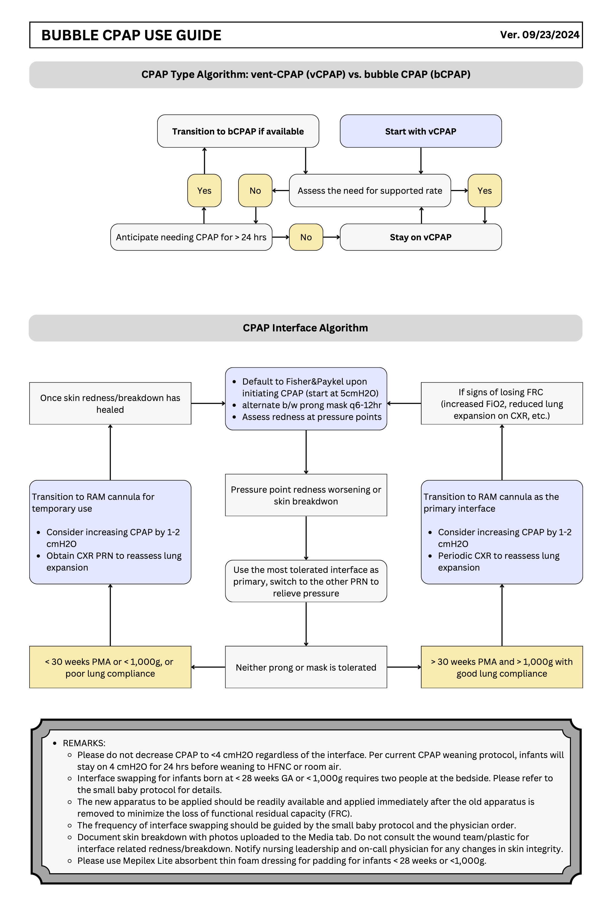
Resources
Section titled “Resources”ELGAN/ELBW Respiratory Support Protocol for the First 72 Hrs
Section titled “ELGAN/ELBW Respiratory Support Protocol for the First 72 Hrs”Protocol
Section titled “Protocol”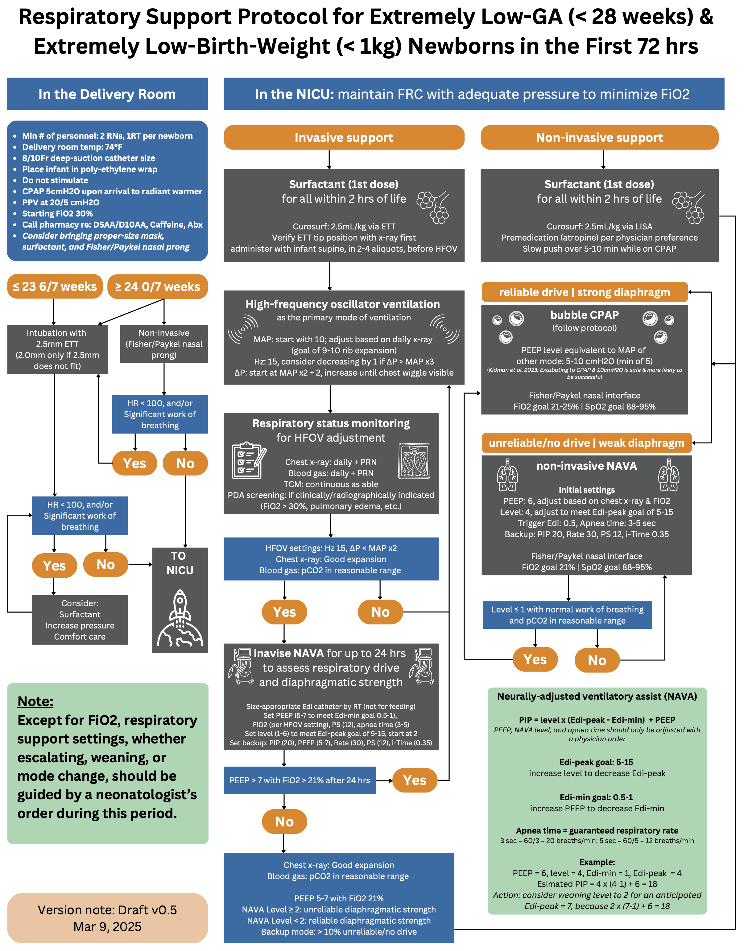
Resource
Section titled “Resource”Neurally adjusted ventilatory assist (NAVA)
Section titled “Neurally adjusted ventilatory assist (NAVA)”NAVA Terminology
Section titled “NAVA Terminology”- Edi: The electrical activity of the diaphragm, considered a respiratory vital sign.
- Edi-peak: The highest value of the Edi signal during a single breath.
- Edi-min: Represents the spontaneous tonic activity of the diaphragm, preventing derecruitment of alveoli during expiration.
- NAVA level: A gain factor converting the Edi signal into proportional pressure.
- Higher NAVA level = more work of breathing provided by the ventilator.
- Lower NAVA level = more work of breathing done by the patient.
Edi Catheter Insertion and Positioning
Section titled “Edi Catheter Insertion and Positioning”- Connect the Edi module and cable.
- Perform the Edi module function check.
- Measure NEX (nose-ear and xiphoid) distance in cm.
- Determine the insertion distance.
- Dip the Edi catheter in water and insert.
- Connect the Edi cable to catheter.
- Verify the position in the catheter positioning screen.
- Check the position of the Edi catheter as a feeding tube according to hospital routines.
- Secure it to the patient, and make a note of the insertion distance.
- Verify the position regularly.
Note: For neonates below 1000 g, use Edi catheter 6 Fr/49 cm.
Positioning Window
Section titled “Positioning Window”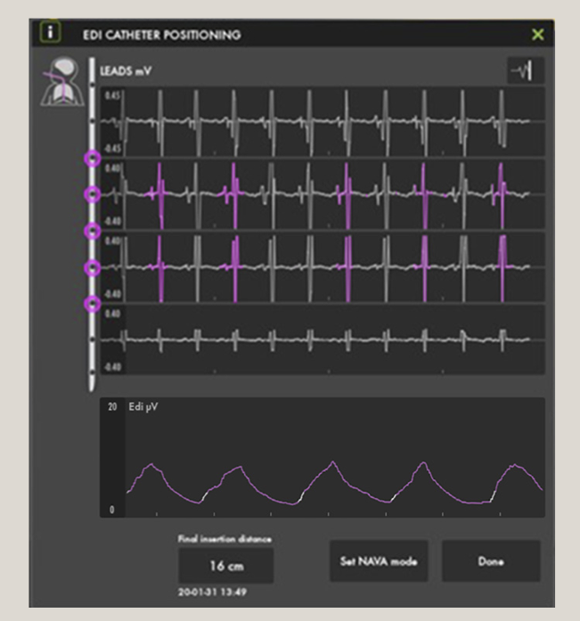
- Correct position:
- Look for a diminishing ECG waveform progressing from the 1st to the 4th waveform.
- Presence of a pink color in the 2nd and 3rd waveforms (may fluctuate to the 1st and 4th waveforms at times).
Re-Positioning
Section titled “Re-Positioning”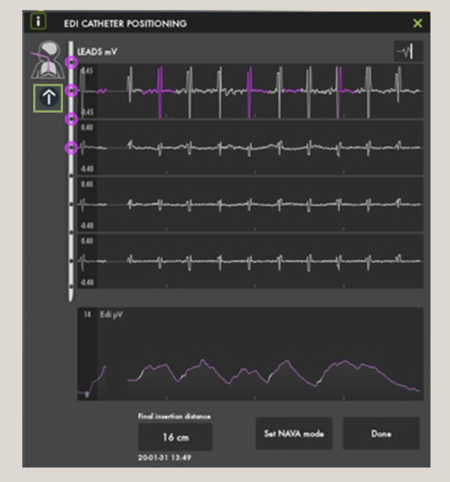
- Too deep:
- Pull out slightly.
- P waves gradually decrease and disappear in the lower leads.
- Dampened QRS.
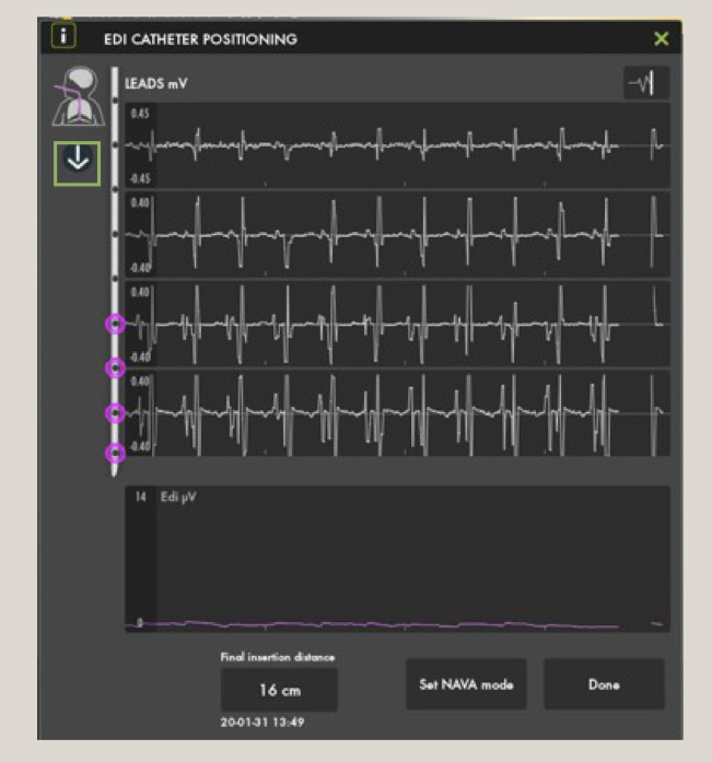
- Too shallow:
- Insert slightly further.
- QRS gets bigger from the top to bottom leads.
Initial NAVA Set-Up
Section titled “Initial NAVA Set-Up”| Parameters | NAVA/NIV NAVA | Management |
|---|---|---|
| NAVA Level | Start with a low NAVA level (1.0-2 cmH2O/μV) | - Titrate to neonates’ comfort and Edi peak 10-15 μV. - If comfortable and Edi peaks <5 μV, decrease NAVA Level in steps of 0.5 until Edi peak values are 5-15 μV. - If increased WOB and Edi peak >20 μV, increase NAVA level in 0.5 increments until comfortable and Edi peaks <15 μV. |
| Edi trigger | 0.5 μV | Avoid “artifact self-triggering” (lower number = more sensitive). |
| Apnea time | 3-5 | Adjust as clinically indicated (minimum rate: 2 seconds = 30 bpm; 1 second = 60 bpm). |
| Ppeak | 35-40 cmH2O NOTE: Pressure limited 5 cmH2O below Ppeak alarm limit (blue alarm). | Set Ppeak high enough for recruiting breaths. Increase if pressure limited alarm is constant. |
| Backup | Activates Pressure Control mode after apnea time if patient is apneic. | Set PC level and RR to assure adequate ventilation. |
NOTE: Set appropriate PEEP for the patient and backup settings: PC above PEEP and RR.
Weaning in NAVA
Section titled “Weaning in NAVA”- Decrease the NAVA level in steps of 0.3-0.5 cmH2O/μV.
- Decrease backup settings if in backup frequently.
- If intubated, extubate to CPAP vs. NIV-NAVA depending on NAVA level, PEEP, and MAP
Troubleshooting
Section titled “Troubleshooting”| Parameter | Investigate the Reason | Management |
|---|---|---|
| High Edi-peak >20 μV | - WOB increased | - Increase NAVA level, increase Ppeak alarm limit |
| - Insufficient backup support | - Increase backup pressure | |
| - Failing NIV treatment | - Intubate and use NAVA | |
| - Discomfort and Pain | - Consider light analgesics | |
| - Edi catheter dislocated | - Reposition Edi catheter | |
| Low Edi-peak <5 μV | - Over-assist | - Decrease NAVA level |
| - Poor respiratory drive | - Decrease backup support | |
| - Sedation too high | - Decrease sedation | |
| Edi-min consistently >5 μV | Atelectasis | - FiO2 high: increase PEEP by 1 - Patient stable: no change |
Additional Information
Section titled “Additional Information”Refer to the SERVO-u/n User’s Manual for operation of the ventilator.
Disclaimer: The assertions stated by physicians are strictly those of the physician and do not necessarily reflect the views of Getinge. Refer to product labeling for current indications, warnings, contraindications, and precautions.
+ MCV00106173 REVA EXP11/22 + MX-7334
Source: Modified from this online document (Click here if the online link is not working).
High-Frequency Jet Ventilator (HFJV)
Section titled “High-Frequency Jet Ventilator (HFJV)”Indications
Section titled “Indications”HFJV is indicated for neonates with any of the following conditions requiring gentle, high-frequency support:
- Pulmonary interstitial emphysema (PIE)
- Respiratory distress syndrome complicated by pneumothorax
- Meconium aspiration syndrome
- Atelectatic pulmonary disease due to secretions or intrapulmonary inflammatory processes
- Respiratory failure (RF) with hemodynamic compromise while on HFOV
- Evolving bronchopulmonary dysplasia (BPD) in extremely immature infants
Note: At the initial roll-out stage, HFJV will not be used as a primary-intention mode of ventilation for extremely preterm infants unless a heterogeneous disease process is evident since birth.
Initial Settings
Section titled “Initial Settings”HFJV operates in tandem with a conventional mechanical ventilator (CMV), which provides PEEP, sigh breaths for recruitment, and bias flow for spontaneous breathing. Use the LifePort ETT adapter for delivery of tiny tidal volumes (<1–2 mL/kg). Start with gentle pressures to avoid injury.
Jet Settings (HFJV - Amplitude/Ventilation for CO₂ Control)
Section titled “Jet Settings (HFJV - Amplitude/Ventilation for CO₂ Control)”- PEEP: Typically 6–10 cmH₂O, or +1 cmH₂O above prior CMV settings; adjust to maintain MAP and oxygenation.
- PIP: Typically starting at 22–24 cmH₂O, may reach high 30’s to low 40’s in immature/sick lungs. PIP controls tidal volume and thus PaCO₂. Due to pressure attenuation in small airways, the lungs only see a small percentage of the set pressure.
Rate (bpm) and I:E Ratio:
| Rate (bpm) | I:E Ratio |
|---|---|
| 420 | 1:6 |
| 360 | 1:7 |
| 300 | 1:9 |
| 240 | 1:12 |
- RR: 240–660 bpm
- 420 bpm for non-extremely preterm or term infants
- 300–360 bpm for extremely preterm infants
- 240 bpm for PIE/air leak
- Rate is chosen based on pathophysiology to optimize I:E ratio and minimize air trapping.
- Note: In HFJV, rate does not affect tidal volume, but may influence minute ventilation and MAP.
- i-Time: 0.02 sec (affects tidal volume). Do not change unless maxed out on PIP and RR and CO₂ remains elevated. Increase by 0.002 sec if necessary to a maximum of 0.03 sec (caution: dramatically affects CO₂ and may cause lung injury).
Sigh Breath Settings (For Lung Recruitment; Contraindicated in Active Air Leaks)
Section titled “Sigh Breath Settings (For Lung Recruitment; Contraindicated in Active Air Leaks)”- PIP: Use low ΔP <5–10 cmH₂O above PEEP for gentle recruitment.
- RR: Typically 0–6 bpm; temporary use only (no more than 30 min if ≥4 bpm) to avoid overdistension. Set to 0 if air leaks are present.
- I-Time: Typically 0.4–0.6 sec; extend for recruitment if needed.
Backup SIMV + PS Support Mode (During HFJV Alarm/Malfunction)
Section titled “Backup SIMV + PS Support Mode (During HFJV Alarm/Malfunction)”Switch to this CMV mode temporarily if HFJV fails:
- PEEP: Match the same setting while on HFJV
- PIP: Typically matching or 2 cmH₂O below HFJV PIP
- RR: 20–40 bpm based on patient needs
- I-Time: 0.3–0.4 sec based on RR
- Pressure Support: 10–12 cmH₂O for 2.5 ETT
Oxygenation Management
Section titled “Oxygenation Management”- Titrate FiO₂ (on both CMV and Jet ventilator) to maintain target SpO₂.
- Consider increasing PEEP/MAP or performing recruitment maneuvers to improve lung volume.
- Notify MD if FiO₂ > 60% acutely.
Additional Initiation Steps
Section titled “Additional Initiation Steps”- Ensure servo pressure stabilizes at 1.5–4.0 psi (auto-adjusts flow to maintain PIP).
- Perform “Rule of 5’s” Recruitment Maneuver if indicated (e.g., for atelectasis): 5 breaths at PIP 5 cmH₂O above PEEP, i-Time 0.5 sec, every 5 minutes. Consider q-shift for ongoing recruitment (avoid in active air leaks).
- Monitor changes in lung dynamics via servo pressure trends.
Management and Titration
Section titled “Management and Titration”Adjust settings based on blood gases, chest X-ray, and clinical status.
Primary goals: - Control CO₂ with Jet PIP/RR - Maintain oxygenation/lung volume with PEEP/MAP - Minimize sigh breaths in air leak-prone conditions
General Titration Guidelines
Section titled “General Titration Guidelines”- CO₂ Management: Increase Jet PIP by 1–2 cmH₂O if PaCO₂ is high; decrease if low. If PIP is maxed, consider lowering RR for air trapping.
- Oxygenation: Adjust PEEP by 1–2 cmH₂O. Use sigh breaths sparingly for recruitment.
- Weaning: Decrease PIP as CO₂ improves; reduce PEEP/FiO₂ as oxygenation stabilizes. Transition back to HFOV when heterogeneous lung pathology is resolved, or to CMV (invasive NAVA) when ready for extubation (to bubble CPAP or non-invasive NAVA).
- Monitoring: Trend servo pressure (rises with decreased compliance/obstruction; falls with leaks/improved compliance). Check ETT position and suction as needed. Obtain ABG 15–30 min after changes.
Disease-Specific Strategies
Section titled “Disease-Specific Strategies”Adapt based on primary diagnosis:
- BPD or evolving BPD: Minimize air trapping/hyperinflation. Use low Jet PIP/RR (e.g., 240–300 bpm), low PEEP, minimal sigh breaths (low ΔP <5 cmH₂O, short i-Time). Consider q-shift Rule of 5’s.
- PIE or Air Leaks: Promote healing. Initiate early; use low Jet RR (240 bpm) for extended expiratory time; optimize PEEP for MAP; avoid sigh breaths.
- MAS: Promote airway clearance via sweeping exhalation. Use low RR; may consider increasing i-Time for term infants; consider sigh breaths.
Troubleshooting
Section titled “Troubleshooting”Use servo pressure trends and alarms to identify issues.
| Issue | Possible Causes | Actions |
|---|---|---|
| Low Servo Pressure (<1.5 psi) | Decreased lung compliance, ETT obstruction/kink, secretions, worsening disease | Suction ETT, check tube position, reassess (X-ray/ABG), decrease PIP if overventilating |
| High Servo Pressure (>4.0 psi) | Improved compliance, air leak, tube malposition, disconnect | Check connections/ETT seal, increase PIP if underventilating, notify MD for potential ETT upsizing |
| High PaCO₂ (Hypercapnia) | Inadequate ventilation, air trapping | Increase Jet PIP by 1–2 cmH₂O; if maxed, decrease RR or increase i-Time (caution); ensure adequate expiratory time |
| Low PaCO₂ (Hypocapnia) | Overventilation | Decrease Jet PIP; increase RR if needed |
| Hypoxemia (High FiO₂) | Atelectasis, low MAP | Increase PEEP by 1 cmH₂O; perform recruitment maneuver (if no air leaks); optimize position |
| Alarms (High/Low Pressure) | Circuit disconnect, obstruction, patient agitation | Inspect circuit/patient box; reset ventilator; switch to backup SIMV+PS if persistent |
| No Improvement/Failure | Wrong rate for pathophysiology, active air leaks | Reassess diagnosis; consider switch to HFOV; notify MD |
Note on HFJV vs HFOV: HFJV has passive exhalation, variable I:E ratio, and low flow. HFOV has active exhalation and fixed I:E ratio.
Dot-phrases
Section titled “Dot-phrases”.JETSETTINGS.JETSETTINGSHINT
Extubation Workflow for ELGAN/ELBW
Section titled “Extubation Workflow for ELGAN/ELBW”Background
Section titled “Background”- Assessing extubation readiness in ELGAN/ELBW babies is challenging.
- Higher extubation failure rates are tolerated in small babies compared to those born at a higher gestational age (GA) or birth weight (BW).
- Early successful extubation correlates with better outcomes, but failure may increase morbidity and mortality.
- Creating a standardized workflow for extubation decision-making and procedures can reduce mental burden for the multidisciplinary care team.
Timing of successful extubation in ELGAN/ELBW from Different Studies
Section titled “Timing of successful extubation in ELGAN/ELBW from Different Studies”| GA | APEX Study | Norway Study | Canada Study | Iowa City, USA | Kanagawa, Japan | Uppsala, Sweden |
|---|---|---|---|---|---|---|
| 22-23 weeks | 28 [27-29.3] | 28 [27-29] | 29.4 [27.4-31.1] | 31 [29-33] | 36.1 [35-37.2] | 29 |
| 24 weeks | 27 [25-29] | 27 [25-29] | 29.1 [26.9-31.0] | N/A | N/A | N/A |
| 25 weeks | 27 [25-29] | 26 [26-28] | 28.1 [25.9-30.3] | N/A | N/A | N/A |
| 26 weeks | 27 [26-29] | N/A | 27.3 [26.3-29.7] | N/A | N/A | N/A |
| 27 weeks | 27 [27-28] | N/A | 27.6 [27.3-29.1] | N/A | N/A | N/A |
Seminars in Fetal and Neonatal Medicine, 2023-10-01, Vol 28, Issue 5, Article 101489
Consensus
Section titled “Consensus”- Adhere to the respiratory protocol for infants on invasive NAVA, testing for up to 24 hours before extubation.
- Extubation decisions require agreement between incoming and outgoing neonatologists.
- Perform extubation in the morning as possible with both neonatologists present in the hospital.
- Ensure at least one neonatologist is present during extubation to evaluate the immediate need for reintubation.
- Keep supplies ready at the bedside.
- Maintain a low threshold for reintubation and restarting HFOV.
- Consider administering a loading dose of caffeine (20 mg/kg) prior to extubation.
Checklist
Section titled “Checklist”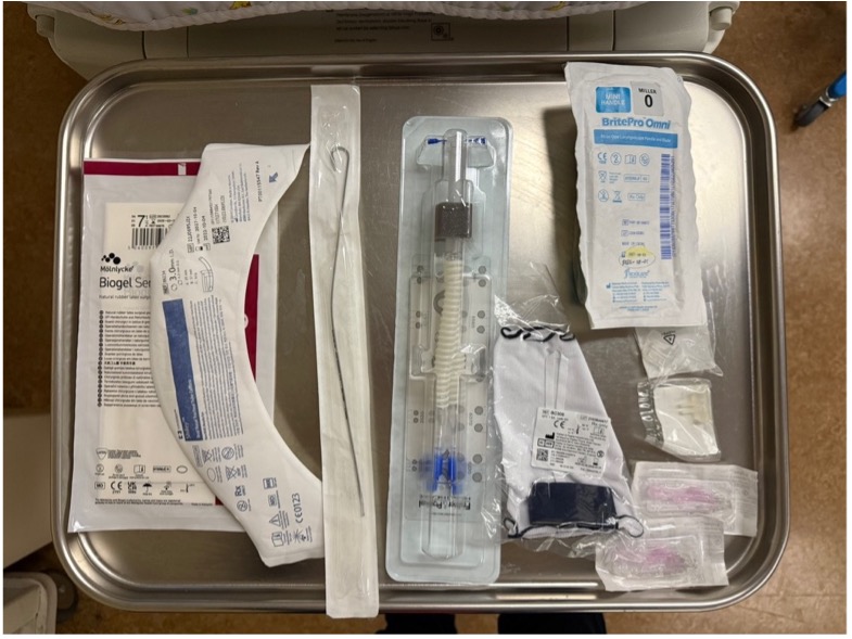
Interface choice
Section titled “Interface choice”- Fisher & Paykel Flexitrunk interface
- Prong / mask
- Bonnet / Tortle cap
even if extubating to Non-Invasive NAVA, Fisher & Paykel interface should still be used, with the goal of weaning to bubble CPAP as soon as the infant is ready
Non-invasive mode selection
Section titled “Non-invasive mode selection”- Bubble CPAP, pressure level: ________
- Non-invasive NAVA (e.g., high MAP, high NAVA level, etc.)
- Edi catheter inserted and depth verified on Servo vent screen
- NAVA and backup settings entered in the Servo machine
- Starting pressure and FiO2
Resuscitation equipment
Section titled “Resuscitation equipment”- T-piece set to 20/5 cmH2O
- ET tube 2.5 and 3.0; stylet
- Laryngoscope size 0 and 00
- Suction catheter 8 and 10 Fr
Medication
Section titled “Medication”- Racemic epinephrine
- Caffeine
Procedures
Section titled “Procedures”-
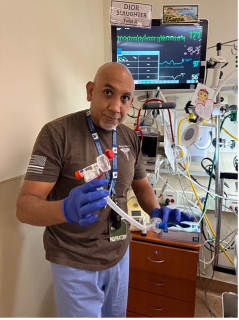
Set up resuscitation equipment
-
Attach non-invasive interface to the infant.
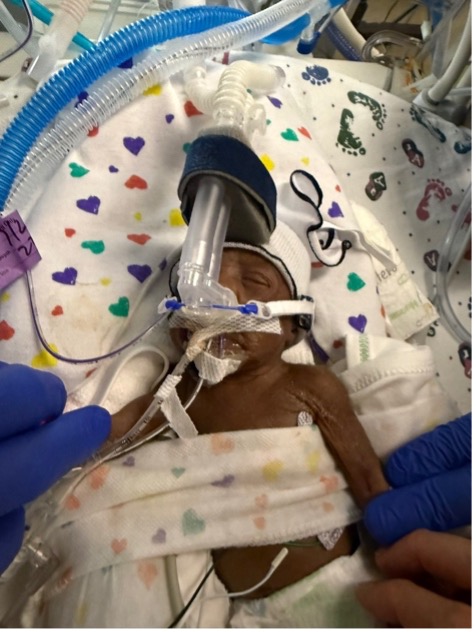
If planning to extubate to bubble CPAP, disconnect one limb first (either incoming or outgoing) before attaching the Flexitrunk and nasal interface to the infant.
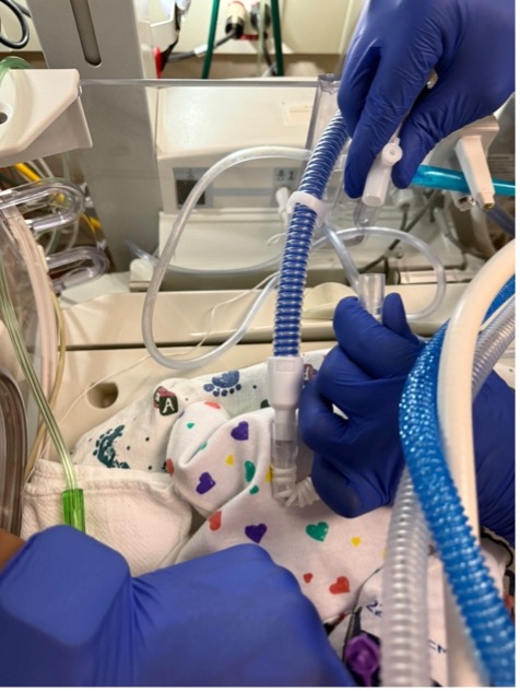
In order for the F/P prong to fit in the nose, ETT tapes may need to be removed first, depending on whether the nostrils are partially obstructed by the tape or not.
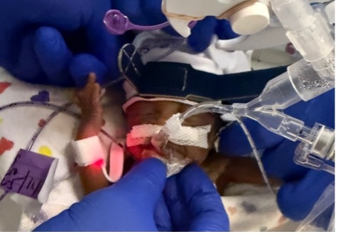
-
Suction the ET tube
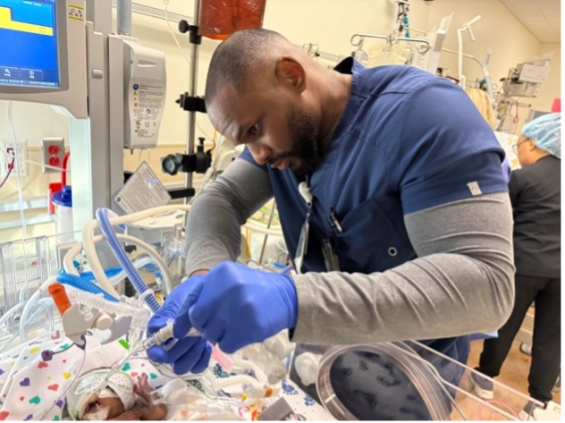
-
Untape and remove the ET tube while connecting the previously disconnected limb to the bubble CPAP device, or switching the vent mode to NON-INVASIVE NAVA
What to expect after extubation
Section titled “What to expect after extubation”Due to fluid shift, the lung fields are expected to white out following extubation for up to 24-48 hrs.
Pre-extubation / Post-extubation Timeline
Section titled “Pre-extubation / Post-extubation Timeline”- Pre-extubation
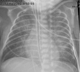
- Post-extubation 12 hrs
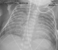
- 24 hrs
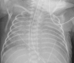
- 36 hrs
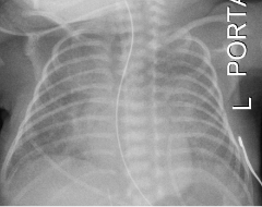
- Four days later
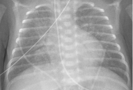
Adjust pressure and FiO2 accordingly to compensate during fluid shift, allowing time to recover.
- Serial chest x-ray and blood gas assessment as needed.
- Fluid management.
- Low threshold to re-intubate if having significant work of breathing and/or respiratory acidosis (e.g., pCO2 > 75, pH < 7.1, etc.)
Cardiopulmonary Events - Spells
Section titled “Cardiopulmonary Events - Spells”Regional Definition
Section titled “Regional Definition”- Pause in breathing for 20 seconds, or
- Pause in breathing with heart rate < 80bpm and/or SpO2 < 80% for ≥ 10 seconds.
Bradycardia
Section titled “Bradycardia”- Does not meeting criteria for Apnea;
- Any drift in HR < 80bpm for ≥ 10 sec requiring intervention other than removing the bottle if during feeding.
Desaturation
Section titled “Desaturation”- Does not meeting criteria for Apnea or Bradycardia;
- SpO2 < 80% for ≥ 10 sec requiring intervention other than removing the bottle if during feeding.
Types of B/D spells
Section titled “Types of B/D spells”Feeding-related spells (feeding spells)
Section titled “Feeding-related spells (feeding spells)”- Apnea or bradycardia occurred during oral feeding and/or up to 30 min after completion of feeding.
Sleep spells
Section titled “Sleep spells”- Spells that are not considered feeding-related.
Bedside Practice Update
Section titled “Bedside Practice Update”Update 1:
Section titled “Update 1:”Do not intervene in the first 10 sec of a spell, unless:
- Life-threatening (e.g. aspiration, airway compromise, etc.).
- Heart rate progress to < 60/min.
Update 2:
Section titled “Update 2:”Do not document events where lowest HR ≥ 80/min and lowest SpO2 ≥ 80% in the flowsheet Apnea/Bradycardia tab:
- These are not A/B/D events by definition.
- If persistent/recurrent SpO2 80-89% and intervention (e.g. stimulation, flow/O2) is needed, please notify MD and document in the notes.
Sleep spells monitoring
Section titled “Sleep spells monitoring”| Requiring intervention | Heart rate | ||
|---|---|---|---|
| ≥ 80/min | < 80/min | ||
| SpO2 | ≥ 80% | Not a spell | 3 days |
| < 80% | 1 day | ||
| Self-resolved | Heart rate | ||
|---|---|---|---|
| ≥ 80/min | < 80/min | ||
| SpO2 | ≥ 80% | Not a spell | No color change: insignificant With color change: 1 day |
| < 80% | No color change: insignificant With color change: 1 day | 1 day | |
Feeding spells monitoring
Section titled “Feeding spells monitoring”| Intervention | Monitoring period |
|---|---|
| Remove bottle only, no color change | insignificant |
| Remove bottle only, with color change | 1 day |
| Stimulation and/or flow/O2 | 1 day |
Spell watch after Hepatitis B vaccine administration
Section titled “Spell watch after Hepatitis B vaccine administration”During spell watch
Section titled “During spell watch”If Hepatitis B vaccine is administered during spell watch, the spell watch period will be modified to include 48 hrs of monitoring following vaccine administration.
- Example: if baby is being monitored from 2/10 to 2/13 (72 hrs), and Hepatitis B vaccine if administered on 2/12, the monitoring period will be extended to 2/14.
Not during spell watch
Section titled “Not during spell watch”No need to monitor for spells after vaccine administration.
Sleep study
Section titled “Sleep study”When an infant is likely to need a sleep study, please contact the following people by starting a Teams group chat:
- Eva Montoya
- Julie Dewitte
- Dennis Hwang
- Huy Truong
- Add any subspecialists for sleep study availability and consultation
Respiratory Support Weaning Algorithm
Section titled “Respiratory Support Weaning Algorithm”flowchart TD
a["<strong>Respiratory support at 32 weeks PMA</strong>"] --> b["<span style='color:red;'>SCENARIO 1:<br>CPAP 4cmH<sub>2</sub>O FiO<sub>2</sub> 21% for > 24hrs</span>"]
a --> c["<span style='color:red;'>SCENARIO 2:<br>CPAP 5cmH<sub>2</sub>O FiO<sub>2</sub> 21%</span>"]
a --> d["<span style='color:red;'>SCENARIO 3:<br>CPAP 5cmH<sub>2</sub>O FiO<sub>2</sub> >21%</span>"]
a --> e["<span style='color:red;'>SCENARIO 4:<br>CPAP >5cmH<sub>2</sub>O</span>"]
c --> b
b --> f("Room air trial")
f --> g["Successful"]
f --> h["Failed"]
g --> j["Room air"]
h --> i("Wean to HFNC 2LPM for 24-72hrs")
i --> p["FiO2<sub>2</sub> remained at 21%"]
i --> q["FiO<sub>2</sub> increased to >21%"]
q --> s["Wean to 21%"]
s --> p
p --> r["Wean flow by 0.25-0.5PM q24-48hrs"]
d --> k("Wean FiO<sub>2</sub> to 21% by 33 weeks PMA")
k --> l["Successful"]
k --> m["Unsuccessful"]
l --> b
m --> o("Consider Diuretics algorithm")
e --> o
q --> o
r --> j
r --> t["Frequent desats during weaning/<br>FiO<sub>2</sub> increased to > 21%"]
t --> u("Consider microflow")
u --> j
o --> v["Able to wean to CPAP 5cmH<sub>2</sub>O FiO<sub>2</sub> 21%<br>by 33 weeks PMA?"]
v --> w["Yes"]
v --> x["No"]
x --> y("Echo for late PH assessment")
w --> cDiuretics Algorithm
Section titled “Diuretics Algorithm”flowchart TD
a["Obtain baseline Chest X-Ray"] --> b("Pulmonary congestion/edema?")
b --> c("Yes")
b --> d("No")
d --> e["Empiric trial of diuretics"]
d --> f["No role of diuretics"]
c --> g["Lasix IV 1mg/kg/day or,<br/> PO 2mg/kg/day for 2 days<br/>(dosing up for discussion)"]
e --> g
g --> h("Improved?")
h --> i("Yes, FiO<sub>2</sub> 21%")
h --> j("Yes, but FiO<sub>2</sub> still >21%")
j --> m["Consider Aldactazide for long-term therapy"]
h --> k("No")
k --> m
k --> l["Consider further workup and/or<br>wean pressure/flow empirically"]
f --> l
l --> p["Consider repeating Chest X-Ray"]
l --> n["Echo for late PH assessment<br/>Especially if frequent desats clinically"]
l --> r["Consider CBC for Anemia"]
i --> o["<strong>Continue to wean<br/>respiratory support</strong>"]
m --> oBronchouplmonary dysplasia (BPD)
Section titled “Bronchouplmonary dysplasia (BPD)”No-BPD prevention bundle (BPD Bundle)
Section titled “No-BPD prevention bundle (BPD Bundle)”General criteria
Section titled “General criteria”- GA < 32 weeks OR BW < 1,500g
Injection meds
Section titled “Injection meds”- Vitamin A
- Reserved for BW < 1,000g AND GA < 28 weeks requiring respiratory support after birth
- Dose: 5,000 units 3 times per week for 12 doses (4 weeks)
- Epogen
- Dose: 250 units/kg/dose SQ Mon Wed Fr starting at DOL7
Enteral meds
Section titled “Enteral meds”When at 100ml/kg/day of enteral feeds:
- Ferrous sulfate 3 mg/kg/dose PO twice daily
- Vitamin E 25 units oral twice daily
Duration
Section titled “Duration”- Discontinue Epogen, Ferrous sulfate, and Vitamin E when infants reach 36 weeks PMA
DART protocol
Section titled “DART protocol”10-day tapering course of dexamethasone sodium phosphate
- 0.15 mg/kg/day for 3 days
- 0.10 mg/kg/day for 3 days
- 0.05 mg/kg/day for 2 days
- 0.02 mg/kg/day for 2 days
Total of 0.89 mg/kg over 10 days
Pneumothorax
Section titled “Pneumothorax”Needle thoractocentesis
Section titled “Needle thoractocentesis”Chest tube placement
Section titled “Chest tube placement”Turkel™
Section titled “Turkel™”- Size: 12-14 Fr
- Suction: -15 cmH2O with the Pleur-evac® Chest Drainage System
Pigtail catheter
Section titled “Pigtail catheter”Video credit: Neoreviews
Modified SELDINGER TECHNIQUE (credit: MacDonald’s Atlas of Procedures in Neonatology)
- GA:
- <28 wks: 6Fr
- >28 wks: 8Fr
- Equipment:
- Introducer and J shaped guidewire
- Finder Needle 20-gauge with syringe
- Dilator
- Adapter for external end of the catheter to connect to suction apparatus.
- Procedures (6 steps):
- Insert finder needle (introducer) on syringe 4th ICS
- Remove the syringe while leaving the finder needle (introducer) & feed the guidewire through introducer (finder needle) to 10cm mark.
- Remove the introducer and finder needle while holding the guidewire steady and insert dilator over the guidewire 1-2cm skin and subcutaneous tissue (not chest wall).
- Remove the dilator and insert the chest tube catheter over the guidewire until all holes are the chest (measures at ~ 7cm).
- Maintain stability of the catheter and remove the guidewire.
- Attach the catheter to the suction drainage device via luer adapter.
- Details and tips:
-
Mount the finder needle syringe with inserting the tip of the needle into the skin at the 4th intercostal space = nipple between the anterior and mid-axillary line above the upper border of the 5th rib.
-
Advance the finder needle slowly, simultaneously applying negative pressure to the syringe. The needle should be advanced perpendicular to the chest wall generally in the direction of the opposite shoulder.
-
Advance the needle until you feel the lack of resistance or “GIVE” and air entry is the syringe.
Do not evacuate more than 1-2mL of air. -
While stabilizing the needle with 1 hand remove the syringe and insert guidewire through the finder needle.
-
Advance the guidewire until the depth marking at 10cm reaches the hub of the needle Advanced the guidewire until 10cm mark that the needle hub
-
Maintain stability of the wire, remove the finder needle.
-
Feed the dilator over the wire and insert in a slow twisting motion to dilate the tract. Advanced the dilator only far enough to dilate the skin tract and parietal pleura. This is normally about 1cm in preterm or 2cm in term infants.
-
Maintaining stability of the wire remove the dilator.
-
Insert the pigtail catheter over the wire and advanced it into the chest wall toward the apex of the lung (anteriorly and medially toward the 2nd intercostal space and midclavicular line) until all the side holes of the catheter are inside the chest wall.
-
This requires uncoiling and holding the pigtail straight with 1 hand while advancing it onto the wire.
-
Maintaining stability of the catheter remove the guidewire. Catheter will spontaneously coil into a pigtail loop inside the pleural space.
-
Attach the luer adapter at the external end of the catheter and connect it the suction apparatus tubing.
-
Secure the chest tube with semipermeable transparent dressing and surgical tape.
-
Suture and the catheter in place is optional but typically not necessary as there is no skin incisions closed and the skin and trach will be snug against the counter.
Warning: it is easy to inadvertently compressed catheter with suture if secured too tightly.
-
Airway Edema
Section titled “Airway Edema”Treatment
Section titled “Treatment”Method 1:
- IV dexamethasone 0.25mg/kg/dose given ~4 hrs prior to scheduled extubastion then q8hrs for a total of 3 doses (Couser 1992).
Method 2:
- IV dexamethasone 0.5mg/kg/dose q8 hrs for 3 doses with last dose administered 1 hr prior to scheduled extubation (Davis 2001).